Official websites use .gov
A .gov website belongs to an official government organization in the United States.
Secure .gov websites use HTTPS
A lock (
) or https:// means you’ve safely connected to the .gov website. Share sensitive information only on official, secure websites.
Taking Measure
Just a Standard Blog
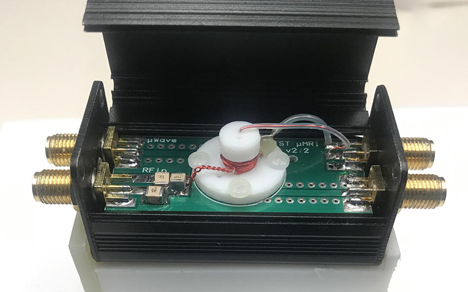
Our microMRI is roughly the size of your hand. The magnet is the red coil that wraps around the sample chamber (white cylinder). The sample is loaded via the tiny hole on the top of sample chamber. This is designed to be the same size as a traditional biopsy needle for point of care testing. On the left and right are connections for the radiofrequency input and output, which power the magnet and hyperpolarization.
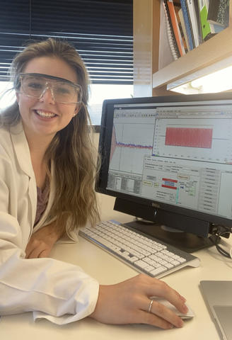
I didn’t really know what to expect on the first day of my virtual Summer Undergraduate Research Fellowship (SURF) program at the National Institute of Standards and Technology (NIST).
At 12:00 p.m., with my second cup of coffee in hand, I sat down at my desk and hopped on the first of many video calls. Brandi Toliver, the SURF director, kicked off the program with introductions of key NIST personnel followed by a presentation about NIST’s history, its aims and its many accomplishments. After this, we broke into small groups and introduced ourselves. One of my peers was an engineer in Georgia; another was interested in cybersecurity and was video-calling from his dorm room in New York. We all had different majors and backgrounds, and were working remotely from different locations. We were all nervous and excited to start our new research projects. It was thrilling to be part of a program with students all over the country!
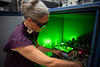
I had opted to work with the Magnetic Imaging Group (MIG), a team with a unique blend of undergraduate students, postdocs and expert scientists. During my time with MIG, my fellow “SURFers” and I learned a little from each of the scientists in the group. Gary Zabow showed us images of tiny micromagnets that expand depending on their environment. This allows scientists to apply color information to black-and-white magnetic resonance imaging (MRI) images to aid in disease detection. Michele Martin talked to us about her single-sided MRI magnets for looking deep underground at the growth of tree roots. With my mentor Stephen Russek, I took my first MRI scans of a human breast "phantom." A phantom is a physical object that is designed to mimic the tissues of the body. Making good phantoms helps clinicians verify their measurements so that they can diagnose diseases like breast cancer with certainty.
Prior to my SURF program, my mentors Stephen Russek and Karl Stupic designed and built a microMRI at NIST. The goal was to make a low-cost tabletop MRI that didn’t need an expensive cooling agent like liquid nitrogen. It could be used by scientists all over the world to grow cells from a tissue biopsy directly in the magnet and image them over time. Although there are current technologies such as microscopes that can image cells, often the sample must be cut, dyed and transferred to a slide, destroying the cells inside. There currently isn’t a good way to grow a cluster of tumor cells and take images to observe them changing over time, for example before and after you administer an anticancer drug. We wanted to solve this problem. My goal for the summer was to optimize the parameters of this microMRI to take higher quality images.
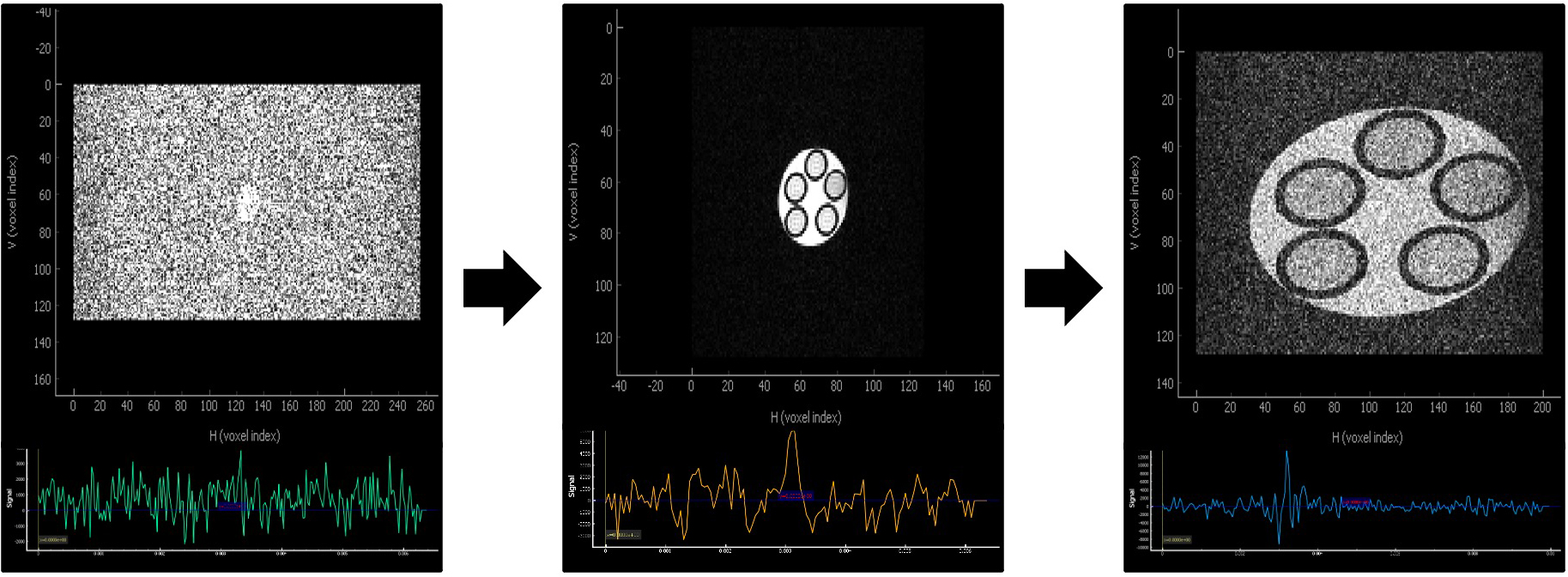
So, what does a virtual research program look like? I began each day by checking in with my mentor and discussing our goals for the day. If it was an imaging day, I would connect remotely to the machine that controlled the microMRI system. I would then acquire images and manipulate the parameters in our pulse sequence to achieve the best resolution possible. It was exceptionally satisfying to observe my images slowly getting better and better. At the same time, I was learning more and more about all the nitty-gritty details behind MRI images that I had taken for granted before. I quickly learned that, like Goldilocks, everything had to be “just right” to get good images.
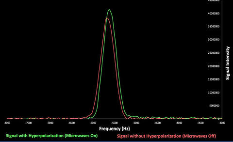
After we optimized the pulse sequence of our microMRI, we worked on the hyperpolarization component of our system. Our tabletop magnet operates using a very small magnetic field (135 milli-tesla), which is about one-tenth of the strength of a typical MRI magnet (1.5 tesla). Because we are using such a small magnetic field, we don’t have as strong a signal as with a larger superconducting magnet in a medical MRI, which results in lower quality images. To combat this, we used hyperpolarized water to increase the signal. Hyperpolarization is a technique where you use microwaves to increase the number of water molecules the MRI can see and increase the overall signal. Other researchers have used hyperpolarization of different compounds like xenon to create high-quality images of the lungs. Just like I had optimized the pulse sequence, I began working on testing out different combinations of microwave power, duration and wavelength to see what boosted our signal the most.
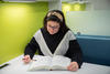
When I received my acceptance to be part of the SURF program, I was finishing up a research semester in a lab at University of Texas Southwestern working on similar hyperpolarization experiments. Since the SURF program was virtual and since both labs worked on similar research, I reached out to both of my project mentors to see if there was an opportunity to bring the two labs together for the summer. We all met and discussed details, and I received permission to spend the day physically at UT Southwestern in Dallas, Texas, while I worked remotely for NIST in Boulder, Colorado.
Over the next few weeks, we figured out ways to work together. We wanted to create a model system to image on the microMRI before we imaged cells. Our model needed to have high contrast, or really light and dark areas that are easy to distinguish from one another. We decided to create liposomes, which are like tiny bubbles with water on the inside protected by an outer coat of lipids (fats). If we put a contrast agent like copper ions on the inside, we could darken the inside of the liposome (copper and water) while keeping the outside bright (fatty lipid coat). We could also make different liposomes with different amounts or concentrations of copper, which would make the inside darker or lighter. I worked on making or synthesizing these liposomes with my group at UT Southwestern.
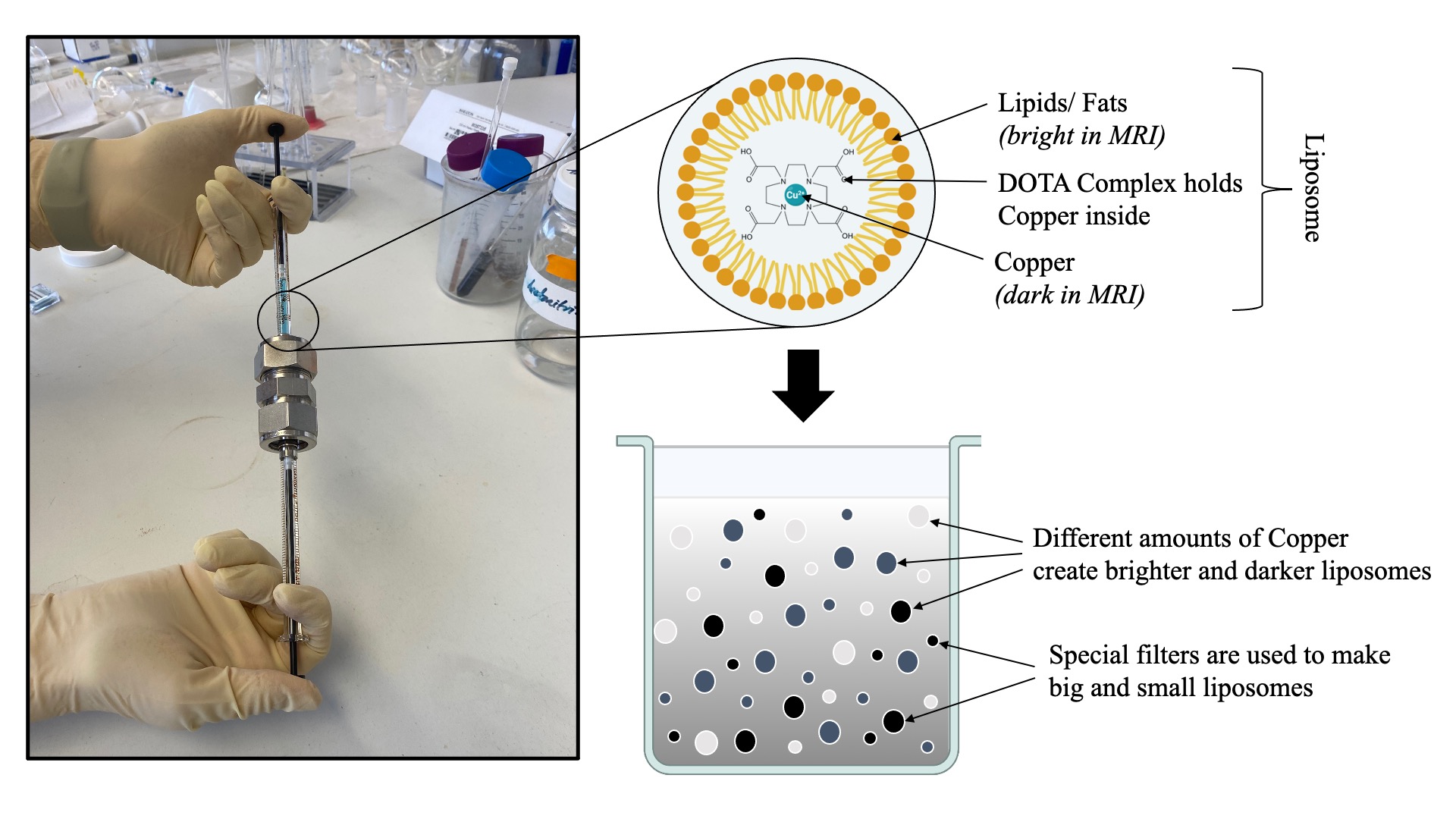
I researched different types of copper-loaded liposomes and pitched the plan to my mentors at UT Southwestern and NIST, who helped me improve my procedure and recommended different laboratory techniques. After we finalized our protocol, I synthesized the liposomes, used a microscope to image them, and sent the results to my NIST mentor. Later my UT Southwestern group became interested in doing low field brain imaging. In the Dallas lab, I made samples and shipped them to Boulder to perform initial imaging experiments at NIST.
My virtual internship allowed me to be in two places at once! As a student, I was able to learn more since each of my mentors had unique experiences and expertise! Furthermore, as many of the MRI conferences transitioned to virtual platforms, I had access to talks and seminars from experts all over the world as a young researcher. As a scientist, collaborating increased the resources I had at my disposal and opened the door for new ideas and experiments. Despite its challenges, remote work during the pandemic has increased opportunities for students like me to collaborate with and learn from people all over the world that would not have been possible otherwise!
Having the opportunity to be part of research as an undergraduate has transformed the way I think about science. It has also helped me better understand the type of work I want to do in the future. I have always loved asking questions and getting my hands dirty, but now I know that I love working on projects to improve diagnostic and imaging technologies. How do we know that dark spot on an MRI image is a tumor? How do we compare results from different MRIs? How do we make images better so that we can make better clinical decisions? These are the kinds of questions I love thinking about and answering. And this is the kind of work that happens at NIST! I am honored to have been a part of the NIST SURF program and am extremely grateful for all the guidance I have received from my mentors. And, I can’t wait for all the exciting projects and questions I will get to tackle as I continue to grow as a scientist!