
Official websites use .gov
A .gov website belongs to an official government organization in the United States.
Secure .gov websites use HTTPS
A lock (
) or https:// means you’ve safely connected to the .gov website. Share sensitive information only on official, secure websites.
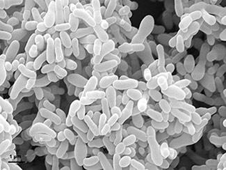
Scanning electron micrograph of Gardnerella vaginalis bacteria
Researchers at the National Institute of Standards and Technology (NIST) and in Lithuania have used a NIST-developed laboratory model of a simplified cell membrane to accurately detect and measure a protein associated with a serious gynecological disease, bacterial vaginosis (BV), at extraordinarily low concentrations. The work illustrates how the artificial membrane could be used to improve disease diagnosis.
Caused by the bacteria Gardnerella vaginalis, BV is a very common health problem in women and has been linked to infertility, adverse pregnancy outcomes, post-surgery infections and increased risk for acquiring sexually transmitted diseases. Current diagnosis relies on time-consuming, labor-intensive and somewhat inconsistent laboratory cultures or immunological assays.
In a recent paper in the journal PLoS One,* researchers at NIST and Vilnius University (Vilnius, Lithuania) reported that they were able to reveal the presence of G. vaginalis by rapidly detecting and quantifying vaginolysin (VLY), a protein toxin produced exclusively by the bacteria, using the NIST model of cell membranes known as a tethered bilayer lipid membrane (tBLM).
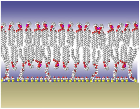
The NIST tBLM is a two-layer sheet of simple lipid molecules analogous to the more complex structures that form the outer shell of animal cells. The membrane is anchored to a substrate with molecular "tethers" that allow it to be surrounded, top and bottom, by typical cellular fluids. Researchers can use the model to study how various factors, such as proteins, affect the integrity of the lipid membrane.
The researchers found that they could detect the presence of VLY down to 28 nanograms (billionths of a gram) per milliliter, a four-fold improvement over antibody detection methods now in use. The speed of detection also is faster, with the tBLM-EIS system yielding results in hours rather than days. Additionally, different G. vaginalis strains produce different amounts of VLY, so in many cases, the corresponding EIS readings can help define the specific type of bacteria present in an infection.
Now that they have proven the viability of the tBLM-EIS detection system, the researchers plan to begin tests on clinical samples early this year.
*R. Budvytyte, M. Pleckaityte, A. Zvirbliene, D.J. Vanderah and G. Valincius. Reconstitution of cholesterol-dependent vaginolysin into tethered phospholipid bilayers: implications for bioanalysis. PLOS ONE (December 2013), DOI:10.1371/journal.pone.0082536.