
Official websites use .gov
A .gov website belongs to an official government organization in the United States.
Secure .gov websites use HTTPS
A lock (
) or https:// means you’ve safely connected to the .gov website. Share sensitive information only on official, secure websites.
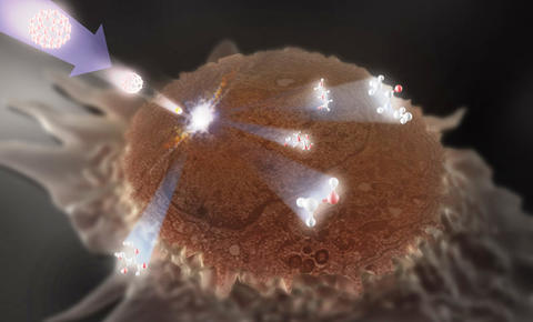
Artist's illustration showing a HeLa cell with its top already "milled" off being probed by a secondary ion mass spectrometry (SIMS) beam. Molecules from three sections of the cell-the membrane, the cytoplasm and the nucleus-are seen ejecting from the surface in response. The spectra from these molecules are used to map the cell sections from which they originate.
To determine if a tissue biopsy reveals the presence of cancer, a histologist often screens for cells with an abnormal appearance or a specific visible trait such as a larger-than-usual nucleus. However, by the time a cancer is physically noticeable, the disease may be in its later stages and more difficult to treat. In an effort to identify the earlier-onset, more subtle chemical changes occurring in a cell heading toward malignancy, researchers at the National Institute of Standards and Technology (NIST) and the National Cancer Institute (NCI) have developed a technique that slices off the top of a cell and makes the structures accessible to spectroscopic examination of their chemical "signature."
Secondary-ion mass spectrometry (SIMS) is a laboratory method developed in the 1960s to define and map the chemicals making up a substance or structure. An ion beam is shot at the surface of a sample, knocking chemical species off the target area that can then be identified by a mass spectrometer. The resulting spectra, in turn, can be used to create a chemical map of the sample.
To date, using SIMS imaging to map mammalian cells has yielded only modest success. To get to the interesting stuff inside the cell, the beam must first blast away the outer cell membrane. Like using a pickax to uncover a fossil, the beam often digs unevenly or too deeply and can damage or destroy the complex molecules and structures inside. The NIST/NCI team tried something more surgical. They first freeze-dried the cell in a manner that prevented its membrane from rupturing and then gently milled the top off the cell with a more powerful, more precisely controlled focused ion beam (FIB) that can skim across the cell at a specified depth. The interior of the cell is left exposed and as close to its natural state as possible for the SIMS beam. "In effect, we get a new, extremely data-rich surface for analysis," says team leader Christopher Szakal.
In a recently published proof-of-concept experiment, the NIST/NCI researchers applied their method to samples from the HeLa immortal human cancer cell line. Specific chemical signals were mapped across the region sliced open by the FIB, yielding images of the cell structures they define at resolutions better than a micrometer (millionth of a meter). For example, spectral maps of phospholipids were used to produce two-dimensional views of cell membranes.
The next step, Szakal says, is to show that the FIB can cleanly slice more than just the top layer off of a cell. "If we can use the FIB-SIMS method to chemically map successive layers of a cell, we'll be able to get three-dimensional images of the cell's components," he explains.
Additionally, the NIST/NCI team is developing mathematical algorithms to enhance and improve the images produced by its new system. The researchers hope that the technique will eventually enable diagnosticians to spot early changes in cell structure that could indicate a move toward abnormality (such as an enlargement of the nuclear membrane) or detect the initial presence of biomarkers, chemical species that can potentially be used to monitor the growth of specific cancers.
C. Szakal, K. Narayan, J. Fu, J. Lefman and S. Subramaniam. Compositional mapping of the surface and interior of mammalian cells at submicrometer resolution. Analytical Chemistry. Vol. 83, Issue 4, pages 1207–1213. Published online Jan. 26, 2011.