
Official websites use .gov
A .gov website belongs to an official government organization in the United States.
Secure .gov websites use HTTPS
A lock (
) or https:// means you’ve safely connected to the .gov website. Share sensitive information only on official, secure websites.
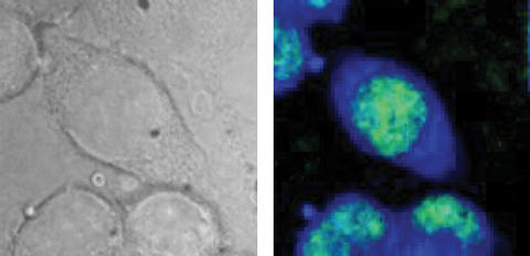
Test cells from a mouse as seen in an optical microscope image (l.), and using B-CARS (r.). The CARS image detects specific molecules to highlight the cell nucleus (green) and intracellular fluid (blue). Images show an area approximately 40 micrometers across. B-CARS image represents approx. 17,000 individual spectra.
*MML
A paper by researchers at the National Institute of Standards and Technology (NIST) may breathe new life into the use of a powerful—but tricky—diagnostic technique for cell biology. The paper, appearing this week in the Biophysical Journal, demonstrates that with improved hardware and better signal processing, a powerful form of molecular vibration spectroscopy can quickly deliver detailed molecular maps of the contents of cells without damaging them. Earlier studies have suggested that to be useful, the technique would need power levels too high for cells.
The technique, "B-CARS,"** is one of several variations on Raman spectroscopy, which measures the frequencies associated with different modes of vibration of atoms and their bonds in a molecule. The exact mix of these frequencies is an extremely discriminating "fingerprint" for any particular molecule, so Raman spectroscopy has been used as a chemical microscope, able to detail the structure of complex objects by mapping the chemical composition at each point in a three-dimensional space.
In the biosciences, according to NIST chemist Marcus Cicerone, Raman spectroscopy has been used to detect microscopic cellular components such as mitochondria, detect how stem cells differentiate into new forms and distinguish between subtly different cell and tissue types. It can, for example, detect minor differences between various precancerous and cancerous cells, potentially providing valuable medical diagnostic information. Even better, it does this without the need to add fluorescent dyes or other chemical tags to identify specific proteins.
The catch, says Cicerone, is speed. The usual method, spontaneous Raman scattering takes a long time to gather enough data to generate a single spectrum—as much as seven minutes for fine detail—and that's for each point in the image. "Seven minutes or even five seconds per spectrum is not feasible when we need a million spectra for an image," he observes. CARS, which uses a pair of lasers to pump up the vibrational states and increase signal, is part of the answer. The current breakthroughs for a broadband CARS instrument developed at NIST since 2004, says Cicerone, gets the same information in 50 milliseconds per pixel.
The new catch is power. Recent papers have argued that to get the necessary data, the lasers used in CARS must run at power levels above the damage threshold for living cells, making the technique nearly useless for clinical purposes. Not quite, according to the NIST team. Their paper describes a combination of improved hardware to gather spectra over a very broad range of wavelengths, and a clever mathematical technique that effectively amplifies the useable signal by examining a portion of signal normally ignored as background interference. The result, says Cicerone, pushes their minimum power level below the damage threshold while retaining the speed of CARS. "We have all the information that you have in a Raman spectrum but we get it 5 to 100 times faster," he says, adding that some obvious modifications should push that higher, opening the door to more widespread use of vibrational spectroscopy in both biology and clinical diagnosis.
** For "broadband coherent anti-Stokes Raman scattering"
S.H. Parekh, Y.J. Lee, K.A. Aamer and M.T. Cicerone. Label-free cellular imaging by broadband coherent anti-Stokes Raman scattering microscopy. Biophysical Journal. V. 99, Oct. 13, 2010.