
Official websites use .gov
A .gov website belongs to an official government organization in the United States.
Secure .gov websites use HTTPS
A lock (
) or https:// means you’ve safely connected to the .gov website. Share sensitive information only on official, secure websites.
Cell membranes form the "skin" of most every cell in your body, but the ability to view them up close and in motion cannot be rendered by many experimental techniques. A team of scientists working at the National Institute of Standards and Technology (NIST) and University of California, Irvine, recently developed a way to magnify them dramatically. Their work has helped illuminate the important role of cholesterol within this boundary between the cell and the outside world.
The multi-institutional team used tools at the NIST Center for Neutron Research (NCNR) to examine the membrane at more than 1,000 times the resolution offered by an optical microscope—the equivalent of magnifying the point of a needle to the size of a large building. This enabled an unprecedented look at the membrane, which—because it controls access to our cells—is a major target for many drugs.
"Drugs that affect pain sensation, heart rhythm, mood, appetite and memory all target proteins lodged in the cell membrane that function like little gates," says Ella Mihailescu of the Institute for Bioscience and Biotechnology Research, a joint institute of NIST and the University of Maryland. "Because membranes and their proteins are important to medicine, we would like a better picture of how the membrane functions—and not just a better snapshot. We want to see it move, as it does constantly in real life."
Optical microscopes offer limited resolution, while the more powerful electron microscopes require freezing samples before they can be magnified. But by using neutron diffraction, which does not require frozen subjects, the team not only observed the membrane more closely and in motion, but they also gained insight into the long-known phenomenon of the membrane growing thicker and stiffer in the presence of cholesterol.
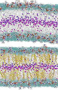
These lipid chains form a two-layer skin with the "heads" of the lipids facing outward toward the cell's exterior and interior and the "tails" intermingling on the inside of the cellular membrane. Cholesterol is known to be important for managing disorder in membranes. The team saw for the first time that when cholesterol is present, these tails line up in a tight formation, looking like a narrow stripe from which the lipid chains stretch outward—and producing the order that had been previously anticipated, but never shown directly. But without cholesterol, the tails go a bit wild, flapping around energetically and in some cases even pushing up toward their chains' heads.
Mihailescu says the findings hint that cholesterol may have profound consequences for the membrane's gatekeeper proteins, which are very sensitive to their environment. "The membrane and its proteins interact constantly, so we're curious to learn more," she says. "With this unique magnification technique, we can explore the cell membrane more effectively than ever possible, and we are now establishing a research program with the University of Maryland to do so in greater detail."
M. Mihailescu, R. G. Vaswani, E. Jardon-Valadez, F. Castro-Roman, J. A. Freites, D. L. Worcester, A. R. Chamberlin, D. J. Tobias and S. H. White. Acyl-chain methyl distributions of liquid-ordered and -disordered membranes. Biophysical Journal, March 2011, Vol. 100, pp. 1455-62, DOI: 10.1016/j.bpj.2011.01.035.