
Official websites use .gov
A .gov website belongs to an official government organization in the United States.
Secure .gov websites use HTTPS
A lock (
) or https:// means you’ve safely connected to the .gov website. Share sensitive information only on official, secure websites.
Summary
The Optical Medical Imaging program was established to advance the application of optical imaging in surgical, clinical, and medical practices. Our goal is develop standards and measurement quality assurance, to characterize and calibrate optical medical imaging technology, and improve surgical and clinical lighting.
Description
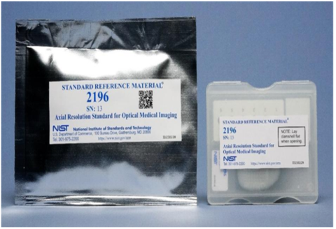
NIST standard reference material (SRM) for the verificaiton of 3D optical medical imaging devices.
We advance measurement science and standards infrastructure to accelerate adoption of optical medical imaging technologies for surgical and clinical applications. Optical medical imaging promises to enhance and complement conventional medical imaging modalities that include magnetic resonance imaging (MRI), computed tomography (CT), and positron emission tomography- computed tomography (PET-CT), which are too expensive for routine applications and too complex for real-time surgical applications. Unlike radiological and x-ray imaging, optical imaging does not expose patients to harmful ionizing radiation and can provide continuous, direct observation throughout protracted surgical procedures as well as long-term patient monitoring for assessing treatment efficacy and progress. Our objectives include enabling SI-traceable calibration and characterization tools for quantitative optical medical imaging technologies, advancing the measurement science for quantitative clinical applications, and enabling the pathway for accelerated Food and Drug Administration (FDA) approval of optical medical imaging devices.
Objectives
- Advance SI-traceable calibration and characterization technology and standards to assess and improve instrument performance through the use of advanced optical tools;
- Capture standard, well-calibrated hyperspectral images of normal and diseased tissues to aid the development of algorithms for quantifying disease and tissue status involving biomarkers;
- Provide the technical foundation to accelerate the certification of optical medical imaging applications by the FDA.
Current Projects
- Photoacoustic Imaging
Photoacoustic imaging is currently the fastest growing medical imaging technology ($1.7B in five years). Commercial systems are just beginning to come on the market and companies are seeking FDA approval in the US for medical imaging applications; however, lack of standards is hampering FDA approvals. Photoacoustic imaging is a hybrid technique combining advantages of optical imaging (high contrast, spatial, and temporal resolution, non-invasive) with ultrasonic imaging (low scattering, deep penetration depth) to provide high-resolution structural, functional, and molecular images deep into biological matter. We are developing SI-traceable photon fluence and resolution phantoms for photoacoustic tomography (PAT) and photoacoustic microscopy (PAM) device characterization and calibration to expedite clinical translation.
- Hyperspectral Imaging for Tumor Margin Analysis
As part of a clinical trial performed at Dartmouth-Hitchcock Medical Center, NIST deployed a calibrated dark-field hyperspectral microscope to image tumor margins in breast lumpectomies. We are analyzing the calibrated hyperspectral scatter data to build libraries of eigenspectra/endmembers spectrally unique to tissue types within an image. We then compare the spatially extracted spectral data with histological annotation by a pathologist to determine normal from cancerous tissue.
- Optical Coherence Tomography
Optical coherence tomography (OCT) is the first FDA-approved optical medical imaging device on the market. We are working to support the clinicians and manufacturers by developing physical standards, known as phantoms, to help calibrate across different manufacturers of OCT systems and across time (to track the progress or treatment of retinal diseases). NIST SI-traceable phantoms will also speed FDA approval for applying OCT to applications beyond retinal imaging, such as the detection of vascular plaques. We have developed a NIST SI-traceable standard reference material (SRM) for the verification of 3D optical medical imaging devices, such as OCT. These SRMs are newly available for sale on the NIST website and through resellers such as Sigma-Aldrich. We are currently developing an advanced SI-traceable retinal eye model with the potential to be included in the NIST Medical Phantom Lending Library.