Taking Measure
Just a Standard Blog

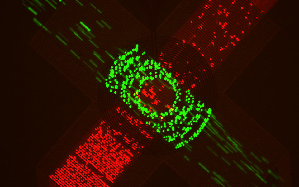
Simultaneous trapping of cancer liver cells (HepG2, red) and endothelial cells (green), each on opposite sites of a porous membrane, for the rapid assembly of tissue/organ on a chip type of microphysiological system. The system has electronic measurement capabilities via integrated electrodes fabricated on the membrane.
It may sound like science fiction, but “organ on a chip” systems — devices capable of imitating the interaction of cells in a specific organ such as the lungs or liver — have begun to be used to test the effectiveness of drugs in the past few years. We use these systems to quantify the physical properties of cells, which provide information regarding the cells’ health before and after treatment with drugs. This is a critical step when determining the usefulness of a therapy.
The methods generally used for this purpose require the cells to be modified with molecules that emit light when exposed to another specific color of light. Differences in the light emitted by cells before and after treatment provide a readout that correlates with the level of the cells’ health. However, light measurements are difficult to reproduce between experiments. In addition, the molecules that are used to make the cells emit light change their interior, producing a somewhat quantifiable but “unnatural” measurement.
At the other end of the spectrum are electronic measurements, which, contrary to cell measurements based on light emission, do not require altering the cells with non-native molecules to determine the cells’ health. These types of measurements take advantage of certain physical properties of cells to distinguish changes in their behavior as a result of exposure to treatments. An electronic device could sense cells’ movements from one place to another, which could provide information on the mobility of metastatic cancer cells, changes in the beating of cardiac cells, and changes in cell shape due to toxins that affect attachment and other internal processes that keep cells alive.
At the National Institute of Standards and Technology (NIST), I have developed and patented a technology that measures physical properties of cells such as their shape, ability to move, and electrical changes between the interior and exterior of the cell. The technology, known as the dual dielectrophoretic membrane for monitoring cell migration, or D2M2CM, can help determine the effects of drugs and other treatments on cells. In addition, with the D2M2CM technique, cells can be placed, or trapped, in specific areas within the measurement device. This particular feature allows for a greater number of cells to be located in the same region, and in turn, provides for a larger number of cells to be analyzed in a shorter time.
While electrical measurements are superior to optical ones in many ways (e.g., label-free, real-time, continuous), today’s biomedical and cell biology basic research communities have had limited experience using electronic measurements, making them less attractive. That’s why I engineered an electronic measurement system that allows for a rapid setup of cells in organ-on-a-chip-type experiments as well as for placing cells specifically on sensors for the measurement of cell migratory behavior. This device has the capability to not only handle cells by trapping them with an electric field, (i.e., dielectrophoresis), but also characterize their physical properties by measuring the electrical resistance of a layer of cells.
This, in combination with other developed components that help cells adhere to the surface, provides for a unique platform that can assess cell migration by measuring the departure of cells from the surface of a porous membrane and, later, their arrival on the opposite side of the membrane after moving through the pores. This can be used as a parameter to determine how aggressive cancer cells are (i.e., metastatic versus nonmetastatic cells). All these measurements can be done in real time, continuously and without the need to label the cells with non-natural molecules that emit light.
Devising a system that can provide electronic measurements in a user-friendly format is the first step toward the use of this technology in, for example, drug testing for cancer and regenerative medicine. My patent was one of two chosen from the NIST portfolio of issued patents or patent applications to participate in the FedTech Spring 2020 Startup Studio. My participation provided me with a better understanding of what customers need in these types of devices and the specific benefits they can get from this technology. As it turns out, I learned that there is a great deal of interest in the capabilities D2M2CM can provide. But customers pointed out that work is still needed in other areas related to the simultaneous detection of multiple parameters, materials utilized as part of the devices to provide biocompatible surfaces for cells and other biological components (e.g., materials used in cell culture dishes such as polystyrene and polycarbonate), and the interface between the user and the instrument for a real-time and easy readout format.
We are continuing research to further develop and transform our technology into a relatively simple method that will allow for the real-time and continuous tracking of cells, thus providing eventual users with a system capable of obtaining quantifiable information on cell behavior and the state of cells’ health.
About the author
Related posts
Comments
Is there a way that you can track the t-cells in multiple myeloma is possibly figure out how to control it. Also is there a study in the works to fix dementia?


were going to a good samaritan in wholeworld