Summary
Neutron detectors based on UV scintillations in xenon gas are under development for a variety of applications. The scintillations are produced with high efficiency by the charged particle products of the neutron capture reaction 10B(n, 𝜶)7Li. Silicon photomultipliers (SiPMs) detect the UV scintillation events that signal a neutron capture event. The Photon Assisted Neutron Detectors (PhAND) are mechanically robust, operate with low input voltages, are scalable, and can be configured in a range of sizes and geometries. Applications include cold neutron detection at the NG-6A beamline at the NIST Center for Neutron Research, neutron detection inside a water phantom at the Maryland Proton Treatment Center, and the reconstruction of neutron source energy spectra with source location.
Description

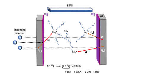
Figure 1. Incident neutrons are absorbed in the 10B(n, 𝜶)7Li reaction. The daughter particles travel out of the boron thin film into a gas volume where they excite xenon to form a short-lived excimer state which releases a far ultraviolet photon upon relaxation. The photon is detected by a silicon photomultiplier. Multiple films are used to create an optical cavity for higher photon detection efficiency. The number of photons produced is a function of the energy deposited by the ⍺ and 7Li which is calculated to be between 103 to 104 photons for a 1 μm boron film.
Due to the simplicity of the PhAND physics package, any number of detector configurations can be deployed. Basic detector operation is illustrated in Fig. 1. Incident neutrons are absorbed in a 10B film and the charged daughter products (𝜶 7Li) enter the surrounding xenon where they produce xenon excimers with high efficiency. Upon decay, the excimer emits far ultraviolet (FUV) radiation centered at 175 nm. The 10B film is electron-beam vapor deposited on thin silicon and aluminum substrates. Film thickness of 1 μm is a compromise between neutron absorption rate and charged particle escape from the film into the xenon gas. On average 103 to 104 FUV photons are produced for every neutron captured in the 10B film. The photons are collected by SiPMs immersed in the gas surrounding the 10B films.
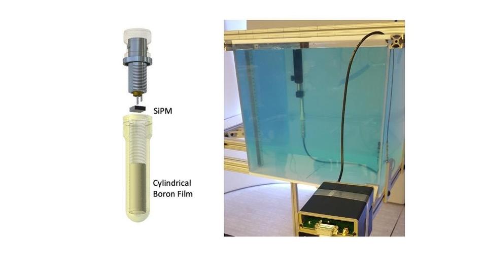
The detectors are mechanically robust, operate with low input voltages, and can be configured in a range of sizes and geometries. Each detector is connected to an electronics processing box that is powered and controlled by USB connection to a computer. A cold neutron beam flux monitor is in operation at the NG-6A beamline with a single planar 10B film. A single film configuration is ideal for beam monitoring while a configuration with 16 films has been used for full beam attenuation. Detection efficiency is directly linked to the number of films used, however the efficiency of detecting an absorbed neutron has been measured to be as high as 75%. Fig. 2 shows a detector with a 10B film deposited on a cylindrical substrate enclosed in a submersible capsule filled with xenon. This detector was used to investigate neutron production in the water phantom at the Maryland Proton Treatment Center.


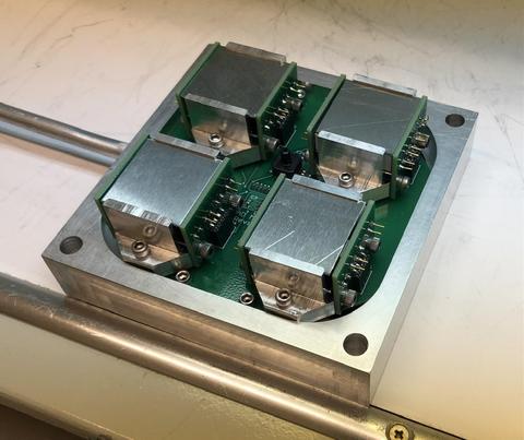
Currently, detectors are being developed to combine into an array to measure neutron energy spectra as the Cellular Array Neutron Spectrometer (CANS). In this work an array of units with different attachments of high-density polyethylene (HDPE) and cadmium make simultaneous measurements of the neutron field. The attachments modify the detector response for each unit in well-determined ways according to the energy of the incident neutrons. The output of the array is fed into machine learning algorithms that reconstruct the neutron source energy spectrum. A unit consisting of four cells is shown in Fig. 3. It consists of four separate 10B film/SiPM cells in a common volume of xenon gas and is one unit of a larger array. One unit is already being used for monitoring neutrons produced by a nuclear fusion experiment at the University of Maryland that requires a strong magnetic field. A full array is also under development for the identification of subsurface water on the Moon through the detection of spallation neutrons diffusing through the lunar regolith. This detector, with a sufficiently advanced neural network and sufficient training data, can even identify the composition of the neutron source material. Current work at the Low Scatter Neutron Calibration Facility is providing the necessary detector response training data, with plans for field testing at the NASA Goddard Geophysical and Astronomical Observatory (GGAO).

