
Official websites use .gov
A .gov website belongs to an official government organization in the United States.
Secure .gov websites use HTTPS
A lock (
) or https:// means you’ve safely connected to the .gov website. Share sensitive information only on official, secure websites.
Summary
A novel neutron lens based on reflection has recently been developed that could greatly benefit neutron imaging and other neutron scattering techniques.
Description
The NIST Neutron Microscope
The NIST neutron microscope based Wolter optics represents a complete change in the way neutron imaging will be conducted, as the time and spatial resolution achievable with this instrument will approach that early generation X-ray synchrotron beamlines. A collaboration between NIST, MIT and NASA is developing the first high spatial resolution neutron microscope based on Wolter optics. The system design suggests that if fully successful, the collaboration will realize spatial resolution of about 3 µm with a time resolution of about 1 s ‒ this about 10,000 shorter exposure time than a conventional pinhole instrument. At the same time, because the sample sits about 1 m from the lens, the neutron microscope will maintain spatial resolution even if the sample is inside a bulky sample environment. The expected increase in the signal rate combined with the improvements in the sample environment will lead to a paradigm shift in neutron imaging. The first full length optics were delivered in summer 2024 and were individually tested at the ICON beamline at the Paul Scherrer Institut while the NCNR recover is underway.
Wolter optics
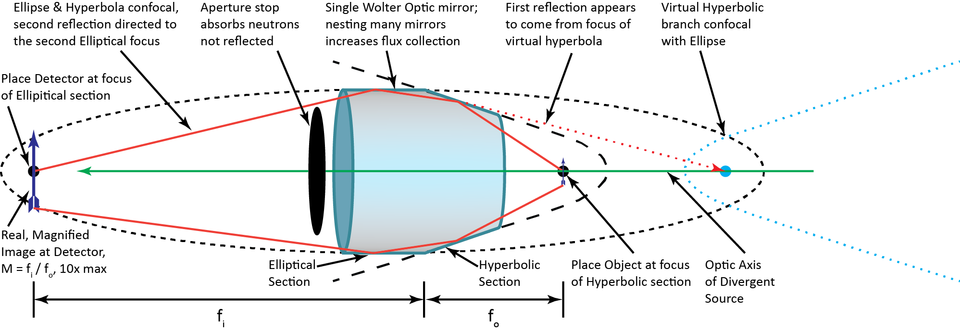
Hans Wolter first proposed using paired, confocal conic section mirrors to act as X-ray lenses in 1952 [1]. The use of two conic sections (including parabolas, hyperbolas and ellipses) eliminates the most severe optical aberrations, such as coma. By pairing a parabola with a hyperbola, one creates a telescope by focusing light coming from infinity and this is a common X-ray telescope configuration including the CHANDRA and soon to launch IXPE telescopes [2]. One can also use the telescope design to create an intense focal spot from collimated radiation such as guided neutron beams. A magnifying imaging objective lens can be realized by pairing an elliptical with a hyperbolic mirror, as shown in the figure below. The focal lengths are measured from the intersection point of the two conic sections. The primary challenge of realizing a neutron microscope based on Wolter optics is creating mirror profiles that deviate from the prescribed angle by less than 10 part per million (10 µradian).
NASA MSFC has been developing such optics for over two decades now for various X-ray astronomy missions and also for laboratory X-ray imaging applications [2], [3]. MSFC utilizes Electroformed Nickel Replication for the fabrication of these lens/mirrors wherein a master mandrel is first made to the desired prescription and surface finish then a thin mirror is electroformed on the mandrel to replicate the mandrel figure. The mirror is then separated from the mandrel using differential thermal expansion, typically in a cold water bath. Several such mirrors of varying diameter but with a common focal length are concentrically nested within a mirror module to increase the collection efficiency. The focus of the research for neutron microscope is to improve the imaging quality of the mirrors by minimizing the figure deviations that are generally imparted during the fabrication process. This overall effort at NASA MSFC will help push the state of art of these grazing incidence neutron mirrors.
Several proof-of-concept demonstrations contributed to the design of the optical system which is expected to arrive in SEP 2022.
First Proof of Concept measurement at NIST
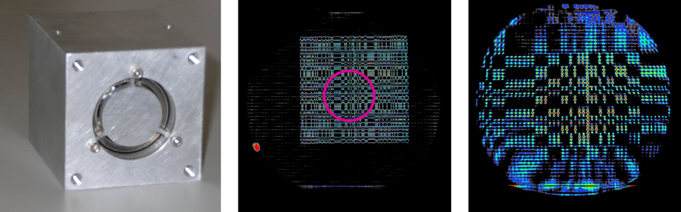
NASA and MIT previously created a prototype optic shown in Figure 1, that consists of the 3 nested coaxial mirror shells with diameters of about 3 cm and an overall length of about 6 cm [4]. The optic has an object focal length of 64 cm and an image focal length of 256 cm so that the optic produces a magnification of 4. A temporary test was installed on the NG1 depth profiling beamline. A grid of 200 µm diameter pinholes in a 100 µm thick Gd foil was imaged using the conventional imaging geometry and then using the prototype Wolter optic [5].
Condenser and Sample Environments
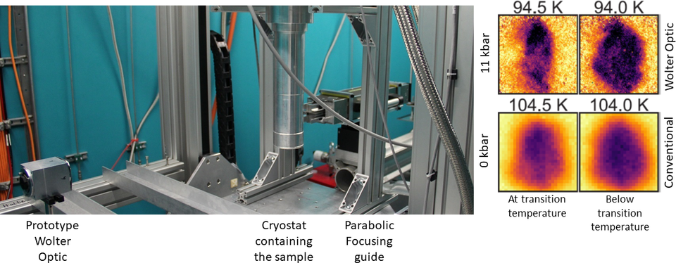
A second proof of concept measurement was conducted in FEB 2017 in collaboration with the imaging team at the FRM II reactor in Garching, Germany using the ANTARES beam line. The goal of the measurement was to image the ferromagnetic transition temperature of a quantum magnet, HgCr2Se4 to determine if the sample underwent a homogeneous transition or there were pressure gradients or compositional inhomogeneities [6]. At atmospheric pressure, the transition temperature is about 104 K and at 15 kbar it is about 91 K. This requires that the sample be placed inside a cryostat and inside a copper-beryllium pressure cell. The transition to the ferromagnetic state is measured by observing the depolarization of the neutron beam, so that a neutron spin filter must be placed between the detector and the sample. The sample environment and spin analyzer are placed about 50 cm between the detector and the sample which, in a conventional neutron imaging setup, results in tremendous image blur. The beam conditioning optics that defines the wavelength and polarizes the beam before the sample creates a tightly collimated beam, which was too collimated to illuminate the 3-cm diameter mirrors and reflect from them. To resolve this, a parabolic focusing mirror was placed just upstream of the sample to create a beam divergence of about 1 degree which optimally illuminated the Wolter optic. In this way, a neutron analog of Hooke’s microscope was realized, with the parabolic mirror acting as a condenser lens and the Wolter optic acting as the objective lens. Using this configuration, which is partially shown in Figure 3, the spatial resolution of this neutron Hooke’s microscope was about 5 times finer than that of the conventional setup. The measurement of the quantum magnet at atmospheric pressure (no applied pressure) was obtained using the conventional imaging setup, while the images of the sample at 11 kbar applied pressure were obtained with the Wolter optic. The process of applying pressure resulted in sample movement that did not enable registering the images.
The NIST Neutron Microscope Based on Wolter optics
The optimized design of the NIST neutron microscope builds on the successful demonstration of a condenser optic with an objective optic. The design also incorporates the upgraded NG6 guides, a spectrum that is composed of longer wavelength neutrons, a 5 m longer flight path, and larger guide cross section that is available to the cold neutron imaging instrument. Based on these upgrades an optimized optical system was designed [7] so that the effective focused flux at the sample position is about 9 × 109 cm-2 s-1. For a conventional neutron imaging system to have a geometric unsharpness of 3 µm with a sample-to-detector separation of 1 cm for a comparable instrument length of the microscope, the effective fluence rate at the sample would be about 5 × 105 cm-2 s-1.
The neutron microscope thus promises to deliver a factor 10,000+ gain in time resolution while yielding current state of the art in spatial resolution. At the same time, with a clear diameter of 1 m around the sample position, the microscope will enable placing samples inside bulky environments such as cryostats, magnets, furnaces, high pressure vessels and rigs without sacrificing the spatial resolution nor require difficult engineering to reduce the distance between the sample and the detector. It is hoped that the microscope optics full potential will be realized with the upgraded CNII.ii after 2027.
References
[1] H. Wolter, “Spiegelsysteme streifenden Einfalls als abbildende Optiken für Röntgenstrahlen,” Ann. Phys., vol. 445, no. 1–2, pp. 94–114, 1952.
[2] B. Ramsey et al., “IXPE mirror module assemblies,” vol. 1111903, no. January 2020, p. 2, 2019.
[3] B. D. Ramsey, “Replicated nickel optics for the hard-x-Ray region,” Exp. Astron., vol. 20, no. 1–3, pp. 85–92, 2005.
[4] B. Khaykovich, M. V. Gubarev, Y. Bagdasarova, B. D. Ramsey, and D. E. Moncton, “From x-ray telescopes to neutron scattering: Using axisymmetric mirrors to focus a neutron beam,” Nucl. Instruments Methods Phys. Res. Sect. A Accel. Spectrometers, Detect. Assoc. Equip., vol. 631, no. 1, pp. 98–104, 2011.
[5] D. Liu et al., “Demonstration of achromatic cold-neutron microscope utilizing axisymmetric focusing mirrors,” Appl. Phys. Lett., vol. 102, no. 18, 2013.
[6] P. Jorba et al., “High-resolution neutron depolarization microscopy of the ferromagnetic transitions in Ni3Al and HgCr2Se4 under pressure,” J. Magn. Magn. Mater., vol. 475, 2019.
[7] D. S. Hussey et al., “Design of a neutron microscope based on Wolter mirrors,” Nucl. Instruments Methods Phys. Res. Sect. A Accel. Spectrometers, Detect. Assoc. Equip., vol. 987, no. October 2020, p. 164813, 2021.