
Official websites use .gov
A .gov website belongs to an official government organization in the United States.
Secure .gov websites use HTTPS
A lock (
) or https:// means you’ve safely connected to the .gov website. Share sensitive information only on official, secure websites.
Cold Neutron Imaging Instrument
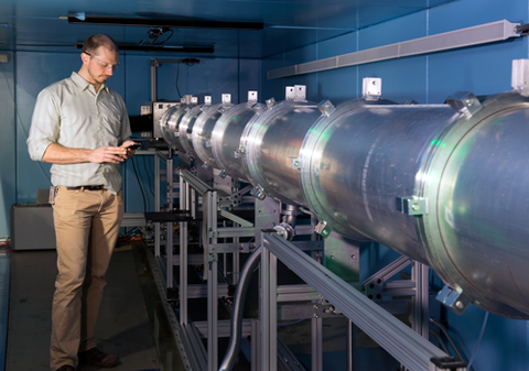
The Cold Neutron Imaging Instrument (CNII) was designed to be a flexible space to test, develop, and employ novel neutron imaging methods to realize different sources of image contrast [1]. The initial motivation for the instrument was to be the home of the first neutron microscope based on Wolter optics. The instrument is capable of polarized neutron imaging, and the first neutron images of an electric field were acquired at the CNII. INFER is a large multi-disciplinary team working to develop dark-field imaging to create multi-scale images that can be thought of as “3D-SANS”. Using a double crystal monochromator, CNII provides users with Bragg-edge imaging to map strain [2] or crystalline phase fractions, for instance in transformation induced plasticity steels [3] or in lead acid batteries. As part of the NCNR cold source upgrade project, the NG6 guide system will be modernized and the new instrument is expected to be available in March 2027.
Beam characteristics
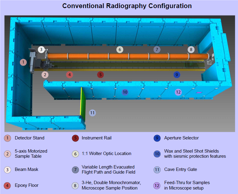
CNII sits at the end position of the neutron guide 6 (NG6). NG6 consists of straight mirrors coated with Ni-58. There is no filter material in the beam which would lower the fluence rate. As a result the neutron beam is an intense cold beam with a thermal equivalent fluence rate (flux) of about 2 × 109 cm2 s-1 at the entrance to the enclosure and downstream 9 m at the sample position it is about 2 × 108 cm2 s1 for a collimation ratio of L/D about 200 with a uniform (wavelength and intensity) area of 10 cm × 10 cm. Variable aperture diameters can be installed to yield larger collimation ratios.
The energy of the beam can be selected either with a double crystal monochromator consisting of highly oriented pyrolytic graphite with mosaic spread of 0.4° (5 cm × 5 cm) or 0.7° (5 cm × 7 cm) or with a neutron velocity selector. The current velocity selector can choose neutron velocities between (1275 m/s and 79 m/s) or alternatively wavelengths between (0.31 nm to 50 nm), with an input beam size of about 6 cm × 10 cm with transmission of about 90% and resolution of about 16%. Vibrations in the rotor exclude wavelengths in the range 0.77 nm to 1.046 nm and 1.52 nm to 2.69 nm. Since the wavelength resolution of the velocity selector is coarser than that of the mosaic crystal (~2 %), the resulting intensity is much greater and finds application in dark-field and phase imaging measurements. The velocity selector will also eliminate artifacts from Bragg edge imaging measurements as it acts as a band pass filter.
Detectors, Scintillators, and Lenses
The CNII makes use of all of the NIST neutron imaging detectors.
Sample manipulation
A wide variety of motorized stages for rotation, tip/tilt, and translation are available to align and manipulate samples during an experiment for complete 6-axis motion. All motorized stages are interfaced with the NIST neutron imaging acquisition software “DataScripting.” Sample environments developed for BT2/NeXT are also available for use at CNII.
Simultaneous dual mode tomography
Similar to the BT2 facility, CNII offers simultaneous neutron and X-ray tomography (“NeXT”). The X-ray generator is a tungsten Bremsstrahlung micro-focus source with maximum electron acceleration of 150 kV and spot size about 7 μm.
Neutron optical components
CNII is designed to allow installation of neutron optical components at any point along the beamline. Components available to users include: a pair of curved neutron polarizers [4], a Talbot-Lau neutron interferometer [5], a suite of transmission and phase gratings to realize 2- and 3-grating far field neutron interferometers [6], [7]. The vacuum tubes that define the flight path are wrapped with wire to create a solenoidal guide field and the tubes can be optionally installed along the flight path per measurement need.
Data Acquisition
Data acquisition is fully automated through using a software package written by the Neutron Imaging Team called Data Scripting.
Data Analysis
Users of the facility have access to both the source code and compiled data analysis packages written in Matlab by members of the Neutron Imaging Team.
References
[1] D. S. Hussey et al., “A New Cold Neutron Imaging Instrument at NIST,” in Physics Procedia, 2015. doi: 10.1016/j.phpro.2015.07.006.
[2] J. W. Sowards, D. S. Hussey, D. L. Jacobson, S. Ream, and P. Williams, “Correlation of Neutron-Based Strain Imaging and Mechanical Behavior of Armor Steel Welds Produced with the Hybrid Laser Arc Welding Process,” Journal of Research of the National Institute of Standards and Technology, vol. 123, no. 123011, pp. 1–8, 2018, doi: 10.6028/jres.123.011.
[3] W. Woo, J. Kim, E.-Y. E.-Y. E. Y. Kim, S.-H. S.-H. H. Choi, V. Em, and D. S. D. S. Hussey, “Multi-scale analyses of constituent phases in a trip-assisted duplex stainless steel by electron backscatter diffraction, in situ neutron diffraction, and energy selective neutron imaging,” Scripta Materialia, vol. 158, pp. 105–109, Jan. 2019, doi: 10.1016/j.scriptamat.2018.08.040.
[4] A. M. Micherdzinska et al., “Polarized neutron beam properties for measuring parity-violating spin rotation in liquid 4He,” Nucl Instrum Methods Phys Res A, vol. 631, no. 1, pp. 80–89, Mar. 2011, doi: 10.1016/j.nima.2010.11.105.
[5] S. W. Lee, D. S. Hussey, D. L. Jacobson, C. M. Sim, and M. Arif, “Development of the grating phase neutron interferometer at a monochromatic beam line,” Nucl. Instrum. Methods Phys. Res., Sect. A, vol. 605, no. 1–2, pp. 16–20, 2009.
[6] D. A. A. Pushin et al., “Far-field interference of a neutron white beam and the applications to noninvasive phase-contrast imaging,” Phys Rev A (Coll Park), vol. 95, no. 4, pp. 1–7, 2017, doi: 10.1103/PhysRevA.95.043637.
[7] D. Sarenac et al., “Three Phase-Grating Moiré Neutron Interferometer for Large Interferometer Area Applications,” Phys Rev Lett, vol. 120, no. 11, p. 113201, 2018, doi: 10.1103/PhysRevLett.120.113201.