Summary
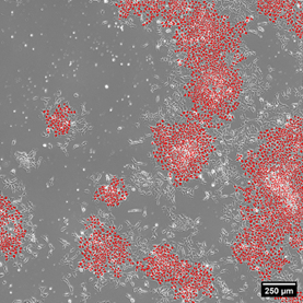
NIST is developing Wide Scale Digital Optical Microscopy (WSDOM) as an advanced measurement capability for imaging induced pluripotent stem cells (iPSCs) and quantifying these live cells as they grow, move, divide and change. With the help of artificial intelligence, hundreds of thousands of individual cells can be imaged and quantified every 2 minutes.
Description
Stem cells are critical starting materials for advanced therapies and diagnostic devices. The most commonly used are induced pluripotent stem cells (iPSCs) which can be created with any individual’s skin cells, and then can be coaxed to become cells for any tissue of the body. Therapies and diagnostics based on iPSCs can therefore be specific for that individual, enabling a truly personalized therapy. The promise of these technologies is great, but measurement challenges exist. WSDOM and the stem cell metrology it enables addresses these measurement challenges in addition to other projects that exist within the Biosystems and Biomaterials (BBD) program in Regenerative Medicine and Advanced Therapy (RMAT), and cell counting, quantitative flow cytometry, and genome editing projects and consortia.
The WSDOM tool images iPSCs at very rapid rates and over long times to quantify hundreds of thousands of individual live cells as they grow, move, divide, and change in their phenotype and gene expression. Each cell behaves a little differently from the others. With the help of artificial intelligence models, individual cells can be identified and quantified every 2 minutes, enabling the tracking of each cell’s characteristics and those of its offspring over time. This work is published.
We are using the temporal, spatial, and gene expression data on each different cell in the population within a theoretical framework based on thermodynamics to provide insight into the biophysical mechanisms by which cells maintain their pluripotency, or alternatively, modify their gene expression to become tissue-specific differentiated cells. This approach to improved metrology will assist the manufacturing and control of stem cells. Because of the complexity of the living cells and their responses to cell culture conditions, it is challenging to know what metrics will provide meaningful assessment of cells, and how to predict which conditions will achieve maximum control over the culture. The new metrics that WSDOM will provide will help to guide the design of manufacturing processes.


